Exam

Comprehensive eye exam

A comprehensive eye exam checks how clearly you see and looks at the health of your whole eye. It can find problems before you notice them.

Key points

How it’s found
An exam can find glaucoma, eye damage from diabetes and other problems before you notice any change in your vision.
Who’s at risk
People with diabetes, high blood pressure, a family history of glaucoma, or contact lenses often need exams more often.
How often
Adults at least every two years, and yearly from age 65. Children before first grade, then yearly.
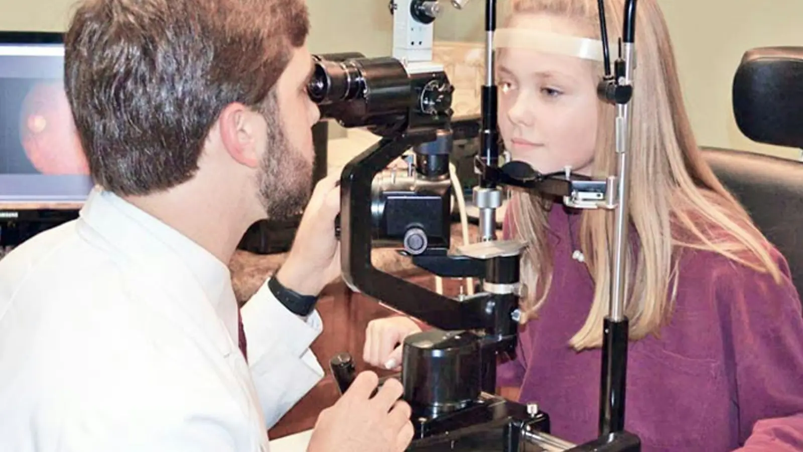
An eye doctor checking a young patient's eyes with a slit lamp microscope

Many eye problems start without any warning signs. Glaucoma, eye disease from diabetes and early macular degeneration often cause no pain and no change in how you see until damage has already begun. A comprehensive eye exam lets your eye doctor find these problems early, when there are more ways to protect your sight.

An eye exam is not the same as a vision screening. A screening at school, at work or for a driver’s license only checks whether you can read letters at a distance. It can miss eye disease, focusing problems and many other issues. A comprehensive exam looks at the whole eye, inside and out.

Your eyes can also show signs of health problems elsewhere in your body, such as high blood pressure, diabetes or high cholesterol. Sometimes an eye exam is the first place these are noticed.

Your eye doctor will start by asking about your eyes, your general health, the medicines you take and whether eye problems run in your family. Tell them how you use your eyes at work, at school and on screens. Then the exam usually includes:

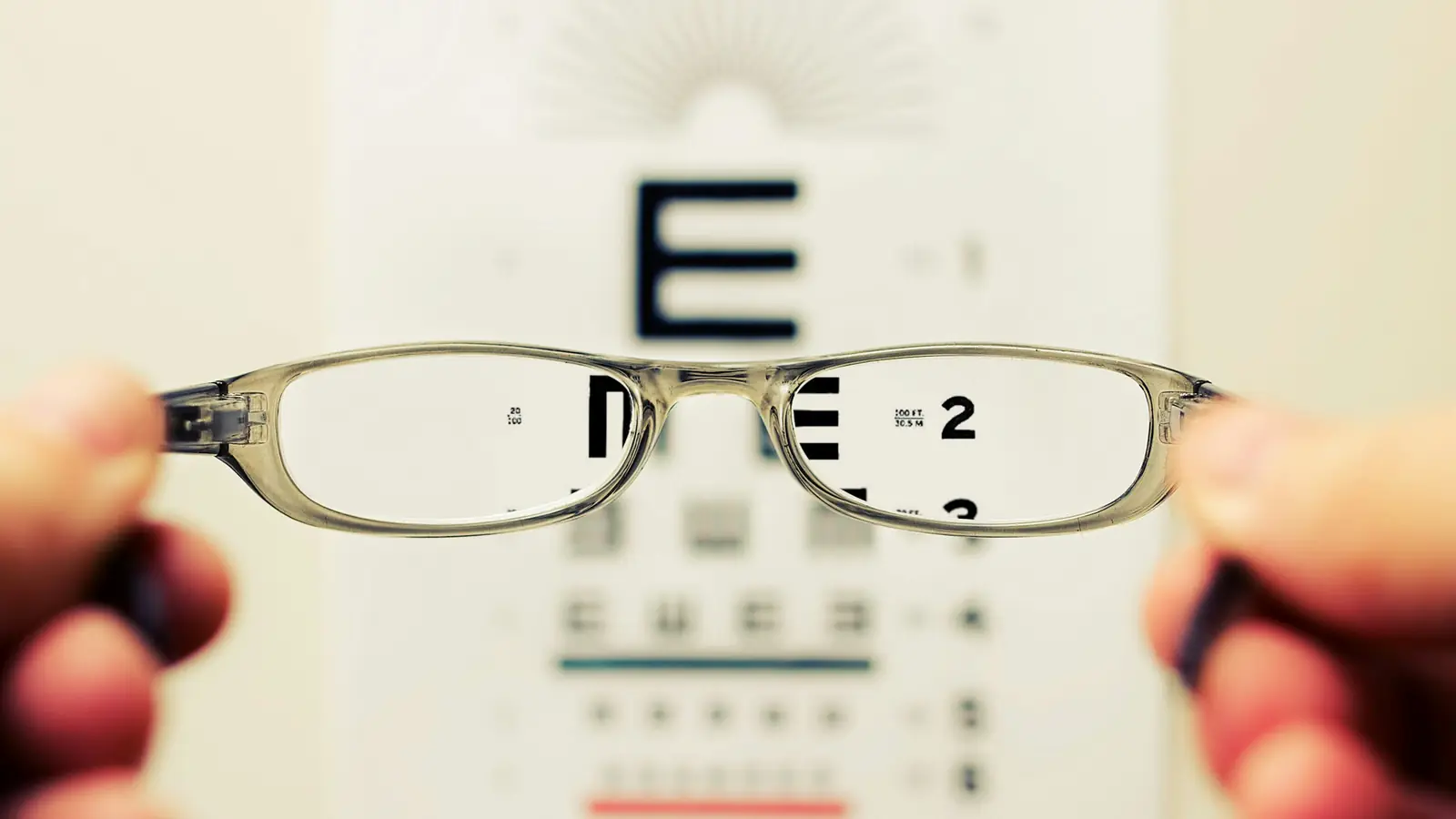
  • Reading an eye chart to measure how clearly you see far away and up close.
  • Checking your prescription to find out whether glasses or contact lenses would help you see better.
  • Testing how your eyes work together, including how they move, focus and team up.
  • Checking your pupils and your side vision.
  • Measuring the pressure inside your eyes, which helps check for glaucoma.
  • Looking at the front of your eye with a special microscope and a bright light.
  • Looking at the back of your eye, including the retina and the optic nerve.

To see the back of the eye well, your eye doctor may use drops that widen (dilate) your pupils. The drops take a little while to work. For a few hours afterward, your vision may be blurry up close and bright light may bother you. Your eye doctor may also take pictures or scans of the back of your eye. These give a record to compare with at your next visit. Pictures can help, but they don’t always replace dilation. Your eye doctor will explain which tests you need and why.

None of the tests hurt. Some eye drops may sting for a moment.